COVID-19 May Invade, Damage Blood Vessels – Not Nerve Cells, says USU Co-Authored Study
By Sarah Marshall
Many individuals with COVID-19 say they experience headaches, along with a “fuzziness” or brain fog that can persist for weeks or month following recovery from the acute respiratory symptoms. This is sometimes referred to as “COVID brain.” The long-term clinical implications of infection by the virus continue to baffle scientists and, until now, the neurological manifestations have been believed to be a result of direct damage to nerve cells. However, a new study in the New England Journal of Medicine suggests the virus might actually damage the brain’s small blood vessels rather than nerve cells, themselves.
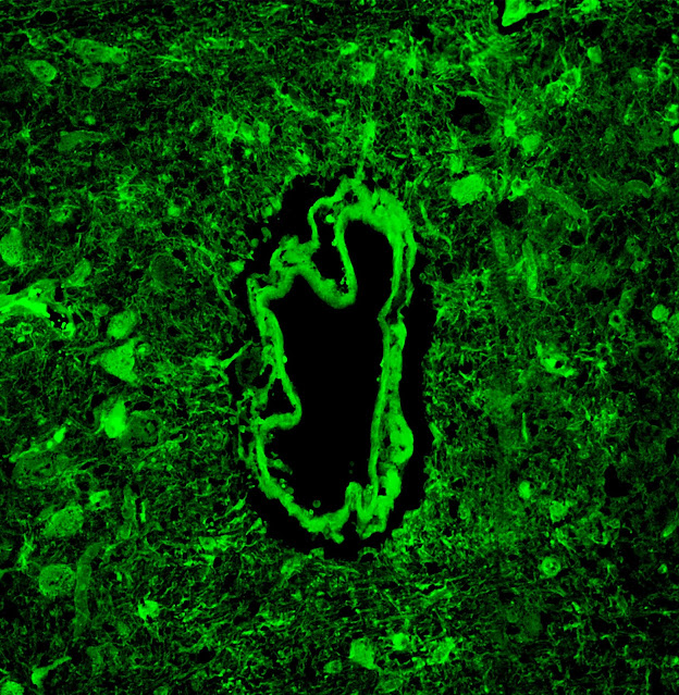
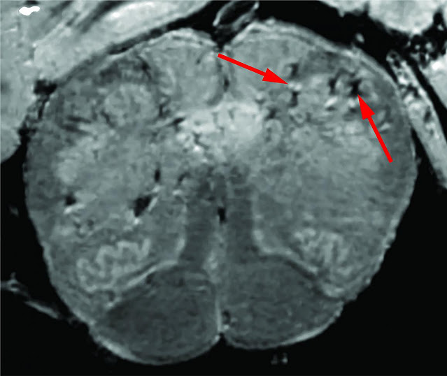
The study, “Microvascular Injury in the Brain of Patients with COVID-19,” was published Dec. 30 and was led by the National Institutes of Health (NIH) and Uniformed Services University (USU) in collaboration with several universities, and other government organizations. Knowing COVID-19 could potentially cause inflammation in and around the blood vessels elsewhere in the body, according to other studies, the researchers took a closer look at the brains of 13 patients who died of COVID-19. High-resolution MRI scanning of brain tissues obtained from these patients, the researchers identified focal hyperintensities, which corresponded to small blood vessels showing evidence of inflammation and damage to their walls. Damage to these vessels were included leakage a protein, fibrinogen, from the blood into adjacent brain regions areas. Despite using very sensitive techniques, the researchers were not able to detect significant levels of SARS-CoV-2, the virus that causes COVID-19, in the brains of the cases they studied – and they found very little evidence of damage to nerve cells.
 |
| High-resolution MRI scanning of brain tissues shows damage to the walls of blood vessels. (NIAID photo) |
Also of significance, the brains of patients studied here were mostly people who died without hospitalization for their disease. Prior studies have mostly looked at patients who endured lengthy hospitalization, including being on ventilators. Such factors may make it difficult to interpret what are the direct effects of the virus on the brain and what are the consequences of lengthy hospital stays on the body, explained study author Dr. Dan Perl, a professor of Pathology at USU and director of the USU Brain Tissue Repository (BTR).
“COVID-19 seems to have a propensity to damage small blood vessels in the brain, rather than the nerve cells themselves,” said Perl.
“While it was tempting to connect our findings of specific brain locations of involvement to specific clinical manifestations, because the cases examined had very little associated clinical information, this could not be done,” said Perl. “These findings address important concepts for understanding the acute and persistent neurologic manifestations of COVID-19, in general. The pandemic is raging in both civilian and military populations and producing considerable mortality and long-term effects among those who have recovered from the infection. The results are of interest and importance to both communities.”
The study was a collaboration between USU, the National Institute of Neurological Disorders and Stroke at the National Institutes of Health, the University of Auckland in New Zealand, the University of Michigan, the Joint Pathology Center, the New York University School of Medicine, and the University of Iowa. To read the full study, visit https://www.nejm.org/doi/full/10.1056/NEJMc2033369.


